Saturday, March 19, 2011
Who Discovered DNA
In 1928, Frederick Griffith performed experiments to prove that DNA carried genetic information. Thereafter, Oswald Avery along with his co-workers identified DNA as the transforming principle in the year 1943. The role of DNA in heredity was confirmed in 1952, when Alfred Hershey and Martha Chase showed that DNA is the genetic material of the T2 phage. Finally in 1953, James D. Watson and Francis Crick suggested the first accurate model of DNA structure. It was the Meselson-Stahl experiment in1958, which led to the final confirmation of the replication mechanism that was implied by the double-helical structure.
The discovery of DNA was one of the major steps in the field of genetics. In case you don't know what DNA stands for, DNA stands for Deoxyribonucleic acid. DNA is a nucleic acid that contains the genetic instructions used in the development and functioning of all known living organisms and some viruses as well. The main function of these DNA molecules is long-term storage of information. In addition to this, if you are looking for more related topics, you can also read about DNA fingerprinting, DNA Research, DNA testing and other related issues like human clothing.
DNA Nanotechnology

DNA nanotechnology uses the unique molecular recognition properties of DNA and other nucleic acids to create self-assembling branched DNA complexes with useful properties. DNA is thus used as a structural material rather than as a carrier of biological information. This has led to the creation of two-dimensional periodic lattices (both tile-based as well as using the "DNA origami" method) as well as three-dimensional structures in the shapes of polyhedra. Nanomechanical devices and algorithmic self-assembly have also been demonstrated, and these DNA structures have been used to template the arrangement of other molecules such as gold nanoparticles and streptavidin proteins.
DNS in Bioinformatics

Bioinformatics involves the manipulation, searching, and data mining of DNA sequence data. The development of techniques to store and search DNA sequences have led to widely applied advances in computer science, especially string searching algorithms, machine learning and database theory. String searching or matching algorithms, which find an occurrence of a sequence of letters inside a larger sequence of letters, were developed to search for specific sequences of nucleotides. In other applications such as text editors, even simple algorithms for this problem usually suffice, but DNA sequences cause these algorithms to exhibit near-worst-case behaviour due to their small number of distinct characters. The related problem of sequence alignment aims to identify homologous sequences and locate the specific mutations that make them distinct. These techniques, especially multiple sequence alignment, are used in studying phylogenetic relationships and protein function. Data sets representing entire genomes' worth of DNA sequences, such as those produced by the Human Genome Project, are difficult to use without annotations, which label the locations of genes and regulatory elements on each chromosome. Regions of DNA sequence that have the characteristic patterns associated with protein- or RNA-coding genes can be identified by gene finding algorithms, which allow researchers to predict the presence of particular gene products in an organism even before they have been isolated experimentally.
DNA in Forensics

Forensic scientists can use DNA in blood, semen, skin, saliva or hair found at a crime scene to identify a matching DNA of an individual, such as a perpetrator. This process is called genetic fingerprinting, or more accurately, DNA profiling. In DNA profiling, the lengths of variable sections of repetitive DNA, such as short tandem repeats and minisatellites, are compared between people. This method is usually an extremely reliable technique for identifying a matching DNA. However, identification can be complicated if the scene is contaminated with DNA from several people. DNA profiling was developed in 1984 by British geneticist Sir Alec Jeffreys, and first used in forensic science to convict Colin Pitchfork in the 1988 Enderby murders case.
People convicted of certain types of crimes may be required to provide a sample of DNA for a database. This has helped investigators solve old cases where only a DNA sample was obtained from the scene. DNA profiling can also be used to identify victims of mass casualty incidents. On the other hand, many convicted people have been released from prison on the basis of DNA techniques, which were not available when a crime had originally been committed.
DNA in Genetic Engineering

Methods have been developed to purify DNA from organisms, such as phenol-chloroform extraction and manipulate it in the laboratory, such as restriction digests and the polymerase chain reaction. Modern biology and biochemistry make intensive use of these techniques in recombinant DNA technology. Recombinant DNA is a man-made DNA sequence that has been assembled from other DNA sequences. They can be transformed into organisms in the form of plasmids or in the appropriate format, by using a viral vector. The genetically modified organisms produced can be used to produce products such as recombinant proteins, used in medical research, or be grown in agriculture.
The History of DNA

The following is a brief history of DNA discovery, analysis, and testing. Highlighted are the significant advances over the last 140 years that evolved into the DNA testing industry and the paternity testing information available today.
1865
The theories of heredity attributed to Gregor Mendel, based on his genetic profiles of pea plants, are well known to any biology student. However, his genetic profiles were so unprecedented at the time, it took 34 years for the rest of the scientific community to catch up. The short monograph, Experiments with Plant Hybrids , in which Mendel described how traits were inherited, has become one of the most enduring and influential publications in the history of science.
1900
The science of genetics was finally born when Mendel's work was rediscovered by three scientists - Hugo DeVries, Erich Von Tschermak, and Carl Correns - each one independently researching scientific literature for precedents to their own "original" work.
1935
Andrei Nikolaevitch Belozersky isolated DNA in the pure state for the first time.
1953
James Watson and Francis Crick proposed the double-stranded, helical, complementary, anti-parallel model for DNA. Nature magazine published James Watson's and Francis Crick's manuscript describing the double helical structure of DNA.
1958
Coenberg discovered and isolated DNA polymerase, which became the first enzyme used to make DNA in a test tube.
1966
The genetic code was "cracked". Marshall Nirenberg, Heinrich Mathaei, and Severo Ochoa demonstrated that a sequence of three nucleotide bases (a codon) determines each of 20 amino acids.
1972
The first successful DNA cloning experiments were performed in California.
1973
For the first time, scientists successfully transferred deoxyribonucleic acid (DNA) from one life form into another. Stanley Cohen and Annie Chang of Stanford University and Herbert Boyer of UCSF "spliced" sections of viral DNA and bacterial DNA with the same restriction enzyme, creating a plasmid with dual antibiotic resistance. They then spliced this recombinant DNA molecule into the DNA of a bacteria, thereby producing the first recombinant DNA organism.
1976
The NIH released the first guidelines for recombinant DNA experimentation. The guidelines restricted many categories of experiments.
1978
Studies by David Botstein and others found that when a restrictive enzyme is applied to DNA from different individuals, the resulting sets of fragments sometimes differ markedly from one person to the next. Such variations in DNA are called restriction fragment length polymorphisms (RFLPs), and they are extremely useful in genetic studies.
1980
Kary Mullis and others at Cetus Corporation in Berkeley, California, invented a technique for multiplying DNA sequences in vitro by, the polymerase chain reaction (PCR). PCR has been called the most revolutionary new technique in molecular biology in the 1980s. Cetus patented the process, and in the summer of 1991 sold the patent to Hoffman-La Roche, Inc. for $300 million.
1984
Alec Jeffreys introduces technique for DNA fingerprinting to identify individuals.
1985
Genetic fingerprinting enters the court room.
1989
Creation of the National Center for Human Genome Research, headed by James Watson, which will oversee the $3 billion U.S. effort to map and sequence all human DNA by 2005.
1990
The Human Genome Project, the international effort to map all of the genes in the human body, was launched. Estimated cost: $13 billion.
1992
The U.S. Army begins collecting blood and tissue samples from all new recruits as part of a "genetic dog tag" program aimed at better identification of soldiers killed in combat.
1993
An international research team, led by Daniel Cohen, of the Center for the Study of Human Polymorphisms in Paris, produces a rough map of all 23 pairs of human chromosomes.
1995
Former football player O.J. Simpson is found not guilty in a high-profile double-murder trial in which PCR and DNA fingerprinting play prominent roles.
1997
Researchers at Scotland's Roslin Institute report that they have cloned a sheep--named Dolly--from the cell of an adult ewe. Polly, the first sheep cloned by nuclear transfer technology bearing a human gene, appears later. Also, leading geneticists expressed shock and dismay as word spread of the US Patent and Trademark Office announcement that it would allow patents on expressed sequence tags (ESTs), short sequences of human DNA that have proven useful in genome mapping.
1998
A rough draft of the human genome map is produced, showing the locations of more than 30,000 genes.
Scientists announce that they have essentially cracked the human genetic code - a decade-long effort by over 1,000 researchers that could revolutionize the diagnosis and treatment of diseases once considered incurable. Decoding the 3 billion chemical "letters" in human DNA is seen as one of history's great scientific milestones - the biological equivalent of the moon landing.
Damage of DNA

DNA can be damaged by many sorts of mutagens, which change the DNA sequence. Mutagens include oxidizing agents, alkylating agents and also high-energy electromagnetic radiation such as ultraviolet light and X-rays. The type of DNA damage produced depends on the type of mutagen. For example, UV light can damage DNA by producing thymine dimers, which are cross-links between pyrimidine bases. On the other hand, oxidants such as free radicals or hydrogen peroxide produce multiple forms of damage, including base modifications, particularly of guanosine, and double-strand breaks. A typical human cell contains about 150,000 bases that have suffered oxidative damage. Of these oxidative lesions, the most dangerous are double-strand breaks, as these are difficult to repair and can produce point mutations, insertions and deletions from the DNA sequence, as well as chromosomal translocations.
Many mutagens fit into the space between two adjacent base pairs, this is called intercalation. Most intercalators are aromatic and planar molecules; examples include ethidium bromide, daunomycin, and doxorubicin. In order for an intercalator to fit between base pairs, the bases must separate, distorting the DNA strands by unwinding of the double helix. This inhibits both transcription and DNA replication, causing toxicity and mutations. As a result, DNA intercalators are often carcinogens, and benzo [a] pyrene diol epoxide, acridines, aflatoxin and ethidium bromide are well-known examples. Nevertheless, due to their ability to inhibit DNA transcription and replication, other similar toxins are also used in chemotherapy to inhibit rapidly growing cancer cells.
Alternate DNA structures

DNA exists in many possible conformations that include A-DNA, B-DNA, and Z-DNA forms, although, only B-DNA and Z-DNA have been directly observed in functional organisms. The conformation that DNA adopts depends on the hydration level, DNA sequence, the amount and direction of supercoiling, chemical modifications of the bases, the type and concentration of metal ions, as well as the presence of polyamines in solution.
The first published reports of A-DNA X-ray diffraction patterns— and also B-DNA used analyses based on Patterson transforms that provided only a limited amount of structural information for oriented fibers of DNA. An alternate analysis was then proposed by Wilkins et al., in 1953, for the in vivo B-DNA X-ray diffraction/scattering patterns of highly hydrated DNA fibers in terms of squares of Bessel functions. In the same journal, James D. Watson and Francis Crick presented their molecular modeling analysis of the DNA X-ray diffraction patterns to suggest that the structure was a double-helix.
Although the `B-DNA form' is most common under the conditions found in cells, it is not a well-defined conformation but a family of related DNA conformations that occur at the high hydration levels present in living cells. Their corresponding X-ray diffraction and scattering patterns are characteristic of molecular paracrystals with a significant degree of disorder.
Compared to B-DNA, the A-DNA form is a wider right-handed spiral, with a shallow, wide minor groove and a narrower, deeper major groove. The A form occurs under non-physiological conditions in partially dehydrated samples of DNA, while in the cell it may be produced in hybrid pairings of DNA and RNA strands, as well as in enzyme-DNA complexes. Segments of DNA where the bases have been chemically modified by methylation may undergo a larger change in conformation and adopt the Z form. Here, the strands turn about the helical axis in a left-handed spiral, the opposite of the more common B form. These unusual structures can be recognized by specific Z-DNA binding proteins and may be involved in the regulation of transcription.
What is mitochondrial DNA?
Although most DNA is packaged in chromosomes within the nucleus, mitochondria also have a small amount of their own DNA. This genetic material is known as mitochondrial DNA or mtDNA.
Mitochondria (illustration) are structures within cells that convert the energy from food into a form that cells can use. Each cell contains hundreds to thousands of mitochondria, which are located in the fluid that surrounds the nucleus (the cytoplasm).
Mitochondria produce energy through a process called oxidative phosphorylation. This process uses oxygen and simple sugars to create adenosine triphosphate (ATP), the cell’s main energy source. A set of enzyme complexes, designated as complexes I-V, carry out oxidative phosphorylation within mitochondria.
In addition to energy production, mitochondria play a role in several other cellular activities. For example, mitochondria help regulate the self-destruction of cells (apoptosis). They are also necessary for the production of substances such as cholesterol and heme (a component of hemoglobin, the molecule that carries oxygen in the blood).
Mitochondrial DNA contains 37 genes, all of which are essential for normal mitochondrial function. Thirteen of these genes provide instructions for making enzymes involved in oxidative phosphorylation. The remaining genes provide instructions for making molecules called transfer RNAs (tRNAs) and ribosomal RNAs (rRNAs), which are chemical cousins of DNA. These types of RNA help assemble protein building blocks (amino acids) into functioning proteins.
What is DNA?

DNA, or deoxyribonucleic acid, is the hereditary material in humans and almost all other organisms. Nearly every cell in a person’s body has the same DNA. Most DNA is located in the cell nucleus (where it is called nuclear DNA), but a small amount of DNA can also be found in the mitochondria (where it is called Mitochondrial DNA or mtDNA).
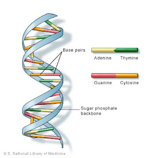
The information in DNA is stored as a code made up of four chemical bases: adenine (A), guanine (G), cytosine (C), and thymine (T). Human DNA consists of about 3 billion bases, and more than 99 percent of those bases are the same in all people. The order, or sequence, of these bases determines the information available for building and maintaining an organism, similar to the way in which letters of the alphabet appear in a certain order to form words and sentences.
DNA bases pair up with each other, A with T and C with G, to form units called base pairs. Each base is also attached to a sugar molecule and a phosphate molecule. Together, a base, sugar, and phosphate are called a nucleotide. Nucleotides are arranged in two long strands that form a spiral called a double helix. The structure of the double helix is somewhat like a ladder, with the base pairs forming the ladder’s rungs and the sugar and phosphate molecules forming the vertical sidepieces of the ladder.
An important property of DNA is that it can replicate, or make copies of itself. Each strand of DNA in the double helix can serve as a pattern for duplicating the sequence of bases. This is critical when cells divide because each new cell needs to have an exact copy of the DNA present in the old cell.